The commonality of statins and Omega 3s
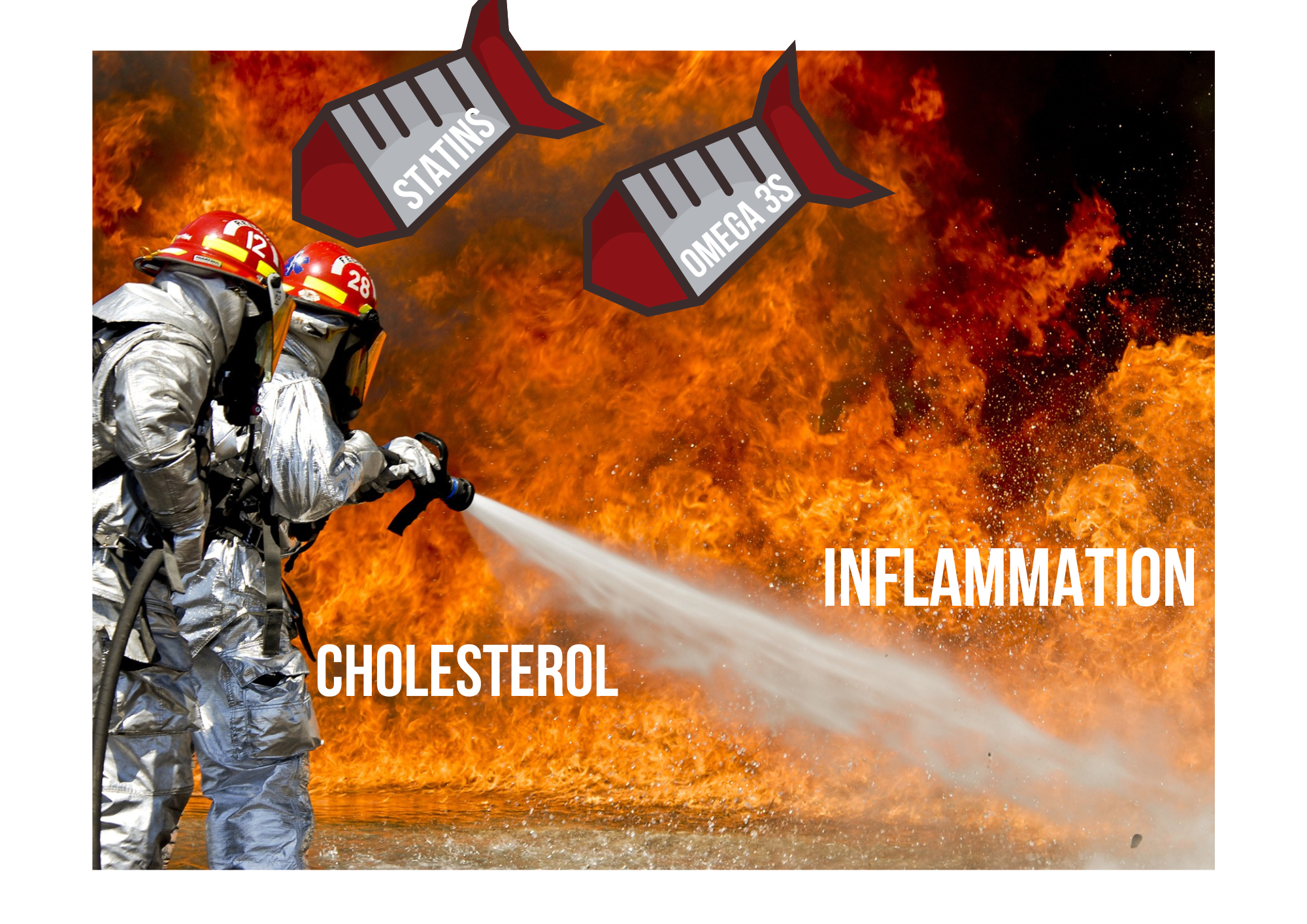
Statins and Omega 3s contribute to long range disorder.
Points to consider –
Increased cholesterol levels appear protective to the central nervous system.
Increasing omega 3s increases aspects of neurological damage.
Statins do not appear to confer any protective effects to the CNS.
Increased PUFA metabolism associated with Parkinson’s and dementia.
Blurring the lines of conferred risk to actual protection.
If the foundation of a house is built on unstable ground, the likelihood of that house staying in its original form is diminished over time. Increasingly the faulty narrative that lipid lowering is heart protective and extrapolated to the central nervous system, muddying the waters further between risk and perceived protection that does not exist. The notion that heart and neurological disease can be prevented by the suppression of lipid levels across a lifetime, ignores the very basics of human physiology.
Historically there’s a vast amount of data and standard textbook descriptions that describe cholesterols antioxidant, anti-inflammatory, and provision of hormones. Two of the main reasons why cholesterol tends to elevate are the well documented factors of hypothyroidism and chronic inflammation. Many people, including MDs still pursue the narrative that cholesterol blocks arteries, leading to heart disease and heart attacks with complete avoidance of the preceding mechanics of such events.
Lowering cholesterol is protective?
Cholesterol is indeed found to block arteries, with an abundance of fatty acids ( a common finding in obese and metabolically compromised is elevated unsaturated fatty acids in cardiac tissue) combined with calcium and it’s the latter that you should be most concerned about. Across ageing and disease, calcium dysregulation occurs and often populates the soft tissues including the arterial walls. Any cardiologist worth their salt (intentional) will often run a coronary artery calcium test to assess how much calcium has permeated the arterial structures (Neves, Andrade, & Monção, 2017). Calcium deposits in these areas are a fundamental feature of a lack of arterial flexibility. Cholesterol tends to aggregate and oxidise around damaged arterial walls in an attempt to repair damage. Perhaps a more appropriate target would be parathyroid hormone, calcium regulation, thyroid metabolism and systemic effects of inflammation?
Since the introduction of statins, there’s been a shift to support the narrative that lowering cholesterol for heart health must also be beneficial for the central nervous system and aspects of neurodegeneration like dementia and Parkinson’s. If you have looked at the vast amount of research and reviews you may have been bamboozled to agree with the papers and mechanistic thinking. Fortunately some reviewers were able to see beyond the bias that exists and failed to locate any protective effects of statins on dementia (Mcguinness, Craig, Bullock, & Passmore, 2016).
PUFA and proposed neurological protection
This tends to be the argument that many people promote and why integrative/ functional medicine doctors fall into the same trap as those recommending statins by suggesting that use of supplements such as Omega 3s, or modulating n3/n6 ratios will lower cholesterol, triglycerides and inflammation. There are many studies that support the notion that lowering cholesterol is associated with worse outcomes in the realms of neurodegeneration (Alecu & Bennett, 2019). Omega 3s degrade cholesterol, disrupt glucose metabolism ( decreased pyruvate dehydrogenase complex) and their accumulation and metabolism in structures in the brain are associated with progression of both Alzheimer’s/dementia and Parkinson’s.
Alpha synuclein (A syn) and its progression to plaque like Lewy body structures share a commonality with dementia and beta amyloid (BA) progression, from functional state (monomer with antimicrobial properties), to misfolded and plaque (Lashuel, Overk, Oueslati, & Masliah, 2013)e like. A syn is a key regulator of synaptic communication, plasticity and maintains nerve function. Just like BA, homogenous targeting and attempting to lower its functional state would be disastrous.
Lashuel 2013
The common feature is increased DHA accumulation, its metabolism and degradation of neuronal structures. However additional insults such as crossing of the blood brain barrier by endotoxin and aluminium exacerbate the negative effects of PUFA metabolism, increased oxidative stress and degradation of the electron transport chain or aerobic metabolism (Exley, 2013; Exley & Mold, 2019; Wang et al., 2018). If dementia is often termed brain diabetes, DHA can be considered a promotor of the diabetic like state in the brain.
DHA is known to induce cell death via increased ceramide production and appears problematic during states of glycolysis (Endo et al., 2018). DHA like many pollutants and estrogens damage optimal aerobic metabolism, creating a vicious cycle that can promote aerobic glycolysis, leading to aggressive utilization of unsaturated fatty acids, often seen in cancer physiology. It shouldn’t be a surprise that increasing levels within the sensitive, glucose preferring areas of the central nervous system respond poorly to oils that are prone to spontaneous oxidation. The metabolic cost of Omega 3s in the brain can only be maintained for so long.
Cholesterol, proteins, water and permeability
The simple narrative of PUFAs being liquid and saturated fats and cholesterol being not so liquid like, still permeates medicine and health. Most saturated fats have a melting point well below 30 degrees centigrade (C) , therefore nearly all oils are in a liquid state in a well heated, healthy mammalian physiology with temperature regulation around 37C. The common notion that membrane fluidity or permeability is regulated by the presence of the unsaturated fats, ignores the concept that saturated fats and cholesterol exhibit both viscous and elastic properties and when depletion of PUFA occurs short chain, saturated fatty acids exhibit the same traits of ‘permeability’( Herman).
Another key aspect in the so called membrane fluidity concept - the most detailed researchers in the field of membrane stability and function, fail to mention the need for PUFA as component of the membrane. Scientists like Gilbert Ling who has provided the most substantial body of work around so-called membrane theory and still (from what I have read, which isn’t always worth much so make your own mind up) remains to be disproven, suggest that phospholipids are a factor, as are proteins and structured water.
It may be added that although in the new model polarized-oriented water has displaced phospholipids as the continuous barrier, phospholipids are nonetheless, important. They may for example, stabilize the protein-water system at the cell surface.In …
It may be added that although in the new model polarized-oriented water has displaced phospholipids as the continuous barrier, phospholipids are nonetheless, important. They may for example, stabilize the protein-water system at the cell surface.
In summary the continuous phase of the cell membrane is polarized oriented water. The most consistently solid component of the cell membrane is protein. The model as such does not require, nor does it exclude, a clear-cut boundary separating the membrane from the cytoplasm like that facing the external medium.
(Ling, 2007)
A concept of optimal biology in the branch of nonequilibrium thermodynamics is that function and structure are best maintained by being far from equilibrium (no energy in-no energy out - therefore staying away from this state, with best working order and optimal energy supply, more complex structures are created). The use of PUFAs as an intervention promote biological decay progressing towards equilibrium. DHA in particular damages the structures associated with optimal function, increases ceramide, uncouples mitochondria, decreases ATP production, and is intimately linked to senescence (Endo et al., 2018).
Saturated fat and cholesterol in the right amount is protective ( much like a variety of food stuffs) and cholesterol does indeed confer stability and protection, despite the suggestion that lowering cholesterol is protective. In a recent study from 2019 the following suggestions were made based upon a follow up study of over twelve million people.
‘ “The lower, the better” cholesterol hypothesis has been accepted by many health professionals. However, the statin trials were mainly performed in persons at a high risk of heart disease, especially in men with manifest CVD, in whom heart disease mortality constituted approximately 50% of all deaths6.
TC ranges associated with the lowest mortality were 210–249 mg/dL in each sex-age subgroup, except for the youngest groups of men, aged 18–34 years (180–219 mg/dL), and women aged 18–34 years (160–199 mg/dL) and 35–44 years (180–219 mg/dL). Inverse associations in the range <200 mg/dL were more than 3-fold stronger than positive associations for cholesterol levels ≥200 mg/dL, except for the youngest adults. Positive associations in the upper TC range were strongest for youngest adults and weakened with advancing age. TC levels <200 mg/dL may not necessarily be a sign of good health.’
(Yi, Yi, & Ohrr, 2019)
A total cholesterol value of approximately 5.5-6.5 mmol/L (210-249 mg/dL appeared the most protective for maintaining health in a substantial amount of people.. If the concept of health is built upon a notion of lowering protective substances, without knowing why those protective substances are elevated; interventions like statins and fish oils only serve to distract from improving biology by managing a symptom. I don’t know about you but to me that’s bonkers.
( For the record I’m not telling anyone taking statins who has defined heart disease to drop the statins and assume all is ok. What I am suggesting is that there are mechanisms beyond a simple good/bad level of cholesterol that require a discussion beyond single system/action medications)
References:
Alecu, I., & Bennett, S. A. L. (2019). Dysregulated lipid metabolism and its role in α-synucleinopathy in Parkinson’s disease. Frontiers in Neuroscience. https://doi.org/10.3389/fnins.2019.00328
Endo, T., Samokhvalov, V., Darwesh, A. M., Khey, K. M. W., El-Sherbeni, A. A., El-Kadi, A. O. S., … Seubert, J. M. (2018). DHA and 19,20-EDP induce lysosomal-proteolytic-dependent cytotoxicity through de novo ceramide production in H9c2 cells with a glycolytic profile. Cell Death Discovery. https://doi.org/10.1038/s41420-018-0090-1
Exley, C. (2013). Human exposure to aluminium. Environmental Sciences: Processes and Impacts. https://doi.org/10.1039/c3em00374d
Exley, C., & Mold, M. J. (2019). Aluminium in human brain tissue: how much is too much? Journal of Biological Inorganic Chemistry. https://doi.org/10.1007/s00775-019-01710-0
Lashuel, H. A., Overk, C. R., Oueslati, A., & Masliah, E. (2013). The many faces of α-synuclein: From structure and toxicity to therapeutic target. Nature Reviews Neuroscience. https://doi.org/10.1038/nrn3406
Ling, G. (2007). History of the membrane (pump) theory of the living cell from its beginning in mid-19th century to its disproof 45 years ago - Though still taught worldwide today as established truth. Physiological Chemistry and Physics and Medical NMR.
Mcguinness, B., Craig, D., Bullock, R., & Passmore, P. (2016). Statins for the prevention of dementia. Cochrane Database of Systematic Reviews. https://doi.org/10.1002/14651858.CD003160.pub3
Neves, P. O., Andrade, J., & Monção, H. (2017). Coronary artery calcium score: current status. Radiologia Brasileira. https://doi.org/10.1590/0100-3984.2015.0235
Wang, L.-M., Wu, Q., Kirk, R. A., Horn, K. P., Ebada Salem, A. H., Hoffman, J. M., … Morton, K. A. (2018). Lipopolysaccharide endotoxemia induces amyloid-β and p-tau formation in the rat brain. American Journal of Nuclear Medicine and Molecular Imaging.
Yi, S. W., Yi, J. J., & Ohrr, H. (2019). Total cholesterol and all-cause mortality by sex and age: a prospective cohort study among 12.8 million adults. Scientific Reports. https://doi.org/10.1038/s41598-018-38461-y